
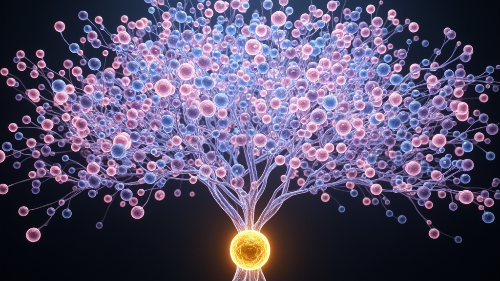
Researchers at the University of Washington School of Medicine and Dartmouth College have used a gene-editing tool called DNA Typewriter to reconstruct the developmental lineage of 1.28 million cells in a mouse embryo over a 13.5-day period, marking the largest cell family tree ever assembled for a mammal.
Tracking 1.28 Million Cells Through Two-Thirds of Mouse Gestation
While researchers mapped the complete cellular lineage of the transparent roundworm decades ago in 1983, doing the same for mammals has remained out of reach because mammalian embryos develop in utero, hidden from direct microscopic view.
The method inserts a synthetic DNA tape into the genome of a fertilized mouse egg. As the embryo grows and cells divide, gene-editing machinery strikes permanent genetic codes onto the tape in strict chronological order.
The study, published October 8, 2026, in the journal Science, followed the recording process across 100 fertilized eggs, yielding 10 embryos suitable for examination. Investigators focused their comprehensive analysis on Embryo No. 3, which successfully gathered the richest developmental history over 13.5 days—representing roughly two-thirds of normal mouse gestation.
How Prime Editing Outperforms Older Cell Tracing Methods
Older cellular tracking techniques often relied on conventional gene-editing tools that cut both strands of DNA. Those older methods frequently left scars, proved harsh on living cells, erased earlier records, and quickly exhausted their recording capacity while forcing scientists to guess timelines afterward.
The DNA Typewriter overcomes these limitations by utilizing prime editing, a next-generation gene-editing technology that cuts only a single strand of DNA. Instead of erasing or deleting genomic sequences, prime editing functions similarly to a word processor that finds a target site, copies information, and pastes it sequentially.
The research team redesigned the synthetic tape so that the accumulated DNA barcodes can be easily retrieved from individual cells later.
When cells divide, the inherited markers are passed down to daughter cells, allowing scientists to sequence individual cell nuclei and reconstruct the exact branching history of the tissue.
Unexpected Discoveries From the First Cell Division
A fortuitous catch occurred right at the very beginning of the experiment, when the fertilized egg split into its first two cells. The recording system marked both founding cells distinctly, creating two independent parallel lineages within the same embryo.
Comparing those two halves revealed an unexpected imbalance in raw cell output. One of the founding branches produced about 57 percent of the sampled cells, while the other accounted for roughly 42 percent.

Yet despite that numerical disparity, both branches generated diverse cell types at nearly equal proportions.
The recorded lineage maps also clarified the schedule of cell fate commitment. Blood cells and retinal cells broke off onto independent developmental paths relatively early, whereas the cells forming the outer layer of skin did not settle into their final identities until much later in the process.
Expanding Applications in Cancer and Organogenesis Research
Beyond charting normal embryonic development, the technology offers new avenues for investigating congenital malformations, genetic disorders, and oncology. By recording cell lineages in high resolution, researchers can pinpoint precisely when and how developmental processes go awry to cause birth defects.
Investigators anticipate that mapping tumor evolution and stem cell differentiation will ultimately assist in the design of therapeutic cells and training data models for virtual embryos.